Different Parts of The Anus and Their Functions with Pictures
Anus Parts:
This 2-inch canal can be distinguished into 9 different parts.
Here is useful information related to anus anatomy.
Two layers of muscles surround this ring-shaped structure. They form the internal anal sphincter and external anal sphincter.
However, both are not under your deliberate action.
The interior sphincter tightens or relaxes under the involuntary action while you can voluntarily control the action of the external one.
The sphincters vary in their length while running along the anal canal.
The internal muscle extends up to 4 centimeters and the external one reaches the maximum size of 10 centimeters or more.
The outer muscular layer retracts during the process of defecation.
Further, it has two strata, namely, deeper stratum and the superficial one (the main portion).
Here follows a brief description of different parts of the anus structure, starting from the anorectal ring and ending at the anal orifice.
- Anorectal Ring
- Anal Canal
- Longitudinal Folds or Anal Columns
- Blood Vessels
- Nerve Endings
- Anus Glands
- Anal Ducts
- Anus Sphincter Muscles
- Anal Valves
Where does the anal region start? What marks the end of rectum and the start of the anal canal?
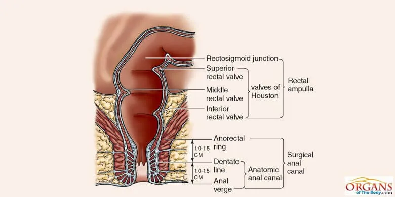
Fig. 5: Anorectal Ring
It is the anorectal ring or the anorectal flexure which serves as a demarcation between the rectum and the anal region.
The anal canal is a small terminal part of the large intestine that starts from the rectum and continues till the anal orifice. It ranges from 3 to 5 centimeters in length.
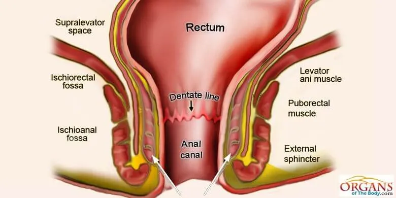
Fig. 6: Anus Canal Diagram.
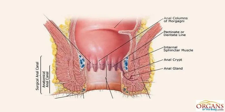
Fig. 7: Anal Columns (formerly Rectal Columns)
The anal canal can be divided into the upper part, lower potion and the anal opening. In the upper part, you will come across the longitudinal folds.
They are also called the anal columns or rectal columns.
There are 5 to 10 rectal columns in the upper part.
The enfolding of the mucous membrane and some of the muscular tissues in the anal canal results in the formation of vertical ridges, called anal columns.
The description of these columns is found in Gray’s Anatomy (1918).
Lots of blood vessels surround the anal canal to supply oxygenated blood to this organ.
In the upper part of anal canal, each rectal column contains a small artery and vein.
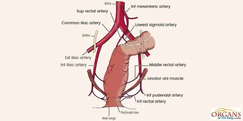
Fig. 8: Blood Vessels in Anorectal Area
Sensitive nerve endings are present in the anal region.
As the waste products enter this region, the nerves take signal to the brain for the generation of an appropriate response.
The anus glands are present in the anal canal of different mammalian species including cats and dogs.
They are also present in humans.
John Aglitis, from the Department of Anatomy at the Ohio State University, published a paper in 1961 discussing the presence and anatomy of the anus glands.
By nature, the anal glands are the eccrine or exocrine glands.
The fluid, secreted by these glands, enters the anal canal via the anal ducts.
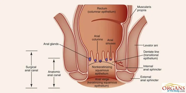
Fig. 9: Location of Anus Glands in the Anal Canal
The wall of the anal canal consists of different layers of muscles, blood vessels, lymph vessels, and sensitive nerve-endings.
Anus glands are also present in this wall at varying lengths.
According to the findings of Eglitis, these glands are vestigial in man.
But still, they have got great clinical significance.
Regarding their size, form, and number, they vary considerably among individuals.
The obstruction of anal ducts can lead to some serious health issues, such as the formation of anus abscesses and anus fistula.
Anal ducts are present in the wall of the anal canal.
The job of these ducts is to empty the secretions of anus glands into the lumen of the canal.
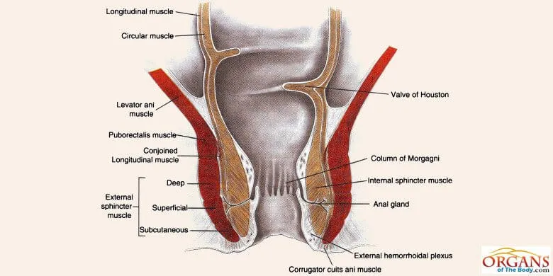
Fig. 10: Anal Ducts in the wall of Anal Canal
A sphincter muscle can also be called an anus ring as it forms a ring-like structure around the anal orifice.
There are two sphincter muscles: internal anal sphincter and external anal sphincter.
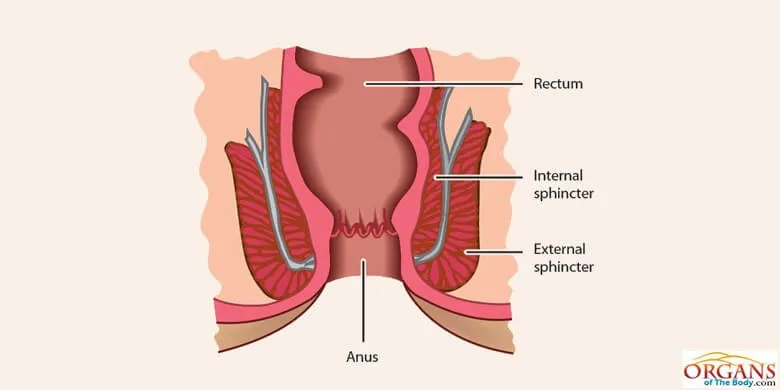
Fig. 11: Internal Anal Sphincter and External Anal Sphincter
The internal sphincter acts involuntarily to regulate the bowel movements.
On the other hand, the external sphincter is under the voluntary control of the individual.
The anal valves are small circular folds of the mucous membrane that join the lower portions of the anal columns.
Small hollow spaces are available between these valves which lead to lymph ducts and the glands.
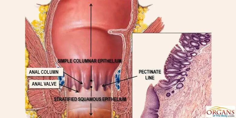
Fig. 12: Anal Valves


